
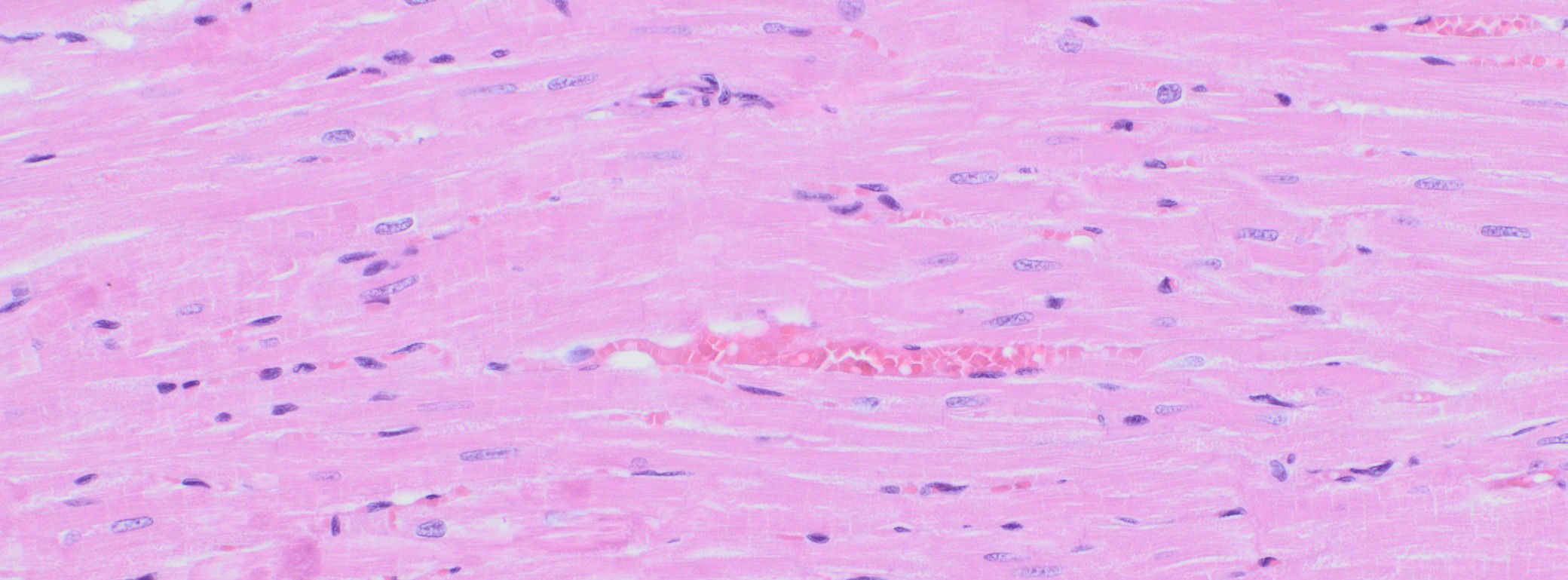
What is Histology?
The study of biological tissue is called histology. It is also known as microanatomy because it involves studying plant, animal and human tissue under a light microscope or an electron microscope. A thin slice of tissue is placed under the microscope and examined after staining. Staining helps in distinguishing various biological structures more easily and accurately. The
What are the Key Stages in Preparing Histology Slides?
There are various techniques that can be used to make histological sections for microscopy. For light microscopes, there are three types of techniques that can be used:
- Paraffin technique
- Frozen sections
- Semithin sections
The most commonly used technique in a histology laboratory is the paraffin technique.
Paraffin Technique
The paraffin technique involves fixing tissues and embedding them in wax to maintain the hardness of the tissues. This makes them easier to slice and examine under a microscope. The process involves the following steps:
- Fixation: Whole organs or spare blocks of tissue are fixed by chemical fixation. The added chemical binds and cross-links some proteins, along with denaturing other proteins by dehydrating them. This process hardens the fixed tissues and prevents degradation by inactivating enzymes. It enhances the staining process and kills bacteria at the same time.
- Dehydration and embedding: After fixation, the tissue is dehydrated and embedded. By dehydrating, all the water from the tissue is removed, which allows it to be embedded in a paraffin solvent called ‘xylene’ as paraffin is not soluble in water. After embedding with paraffin, the sample will solidify.
- Sectioning: After the tissue is solidified, it is trimmed and mounted on a microtome, which gives a cut at around 100 μm. The cut is stained and then put on a slide. The cut needs also colored to be able to distinguish different tissue types and cells. There are several staining methods. The choice of staining depends upon what needs to be investigated and examined.The colored section is studied with advanced histological technology and from these samples written finally a report. The process may look different depending on what type of study it is, something that also applies to the process time.
Frozen Sections
In this method, tissues are frozen with liquid nitrogen. They are then cut in a refrigerated cabinet, also known as a
Semithin Sections
The sections are embedded in epoxy or acrylic resin to create sections that are thinner than less than 2 µm. This is done because it is sometimes hard to see details in thicker sections.
What are the Uses of Histology?
Histology is used to investigate various types of tissues. A histology CRO examines the contents of the tissue. Histology can also be used to investigate agricultural land, for example, in order to observe chemicals that can be found in the soil. Histology is also used for autopsies.
What are Some Other Histology Services?
There are many histology services, including neuropathological histology. This field investigates nerves to find diseases, nerve damage and the like. A histological examination can confirm a diagnosis.
The histology process for these types of studies differs from the routine histology. Other examples of histology services are:
- Autopsy
- Histology of special species (fish, birds, etc.)
- Plastic injection for neurotoxic studies
- Histological and toxicological pathology
- Reproductive and fetal pathology
- Molecular pathology