AnaPath keeps expanding its expertise by offering in-house scanning electron microscopy (SEM) equipped with energy-dispersive X-ray spectroscopy (EDX) (syn. EDS, EDXS or XEDS) services.
Therefore, with a magnification rate of 200-1,000,000 x and 2.5nm resolution, we can analyse biological samples and inorganic objects at nanoscale sizes, as well as determining their chemical composition. Biological samples such as fresh, frozen or dried organs/tissues and tissue sections can be directly analyzed on our SEM/EDX instrument.
Additionally, in order to improve quality of the images for low conductive samples, AnaPath also offers the possibility to platinum-coating the sample before SEM/EDX analysis.
SEM
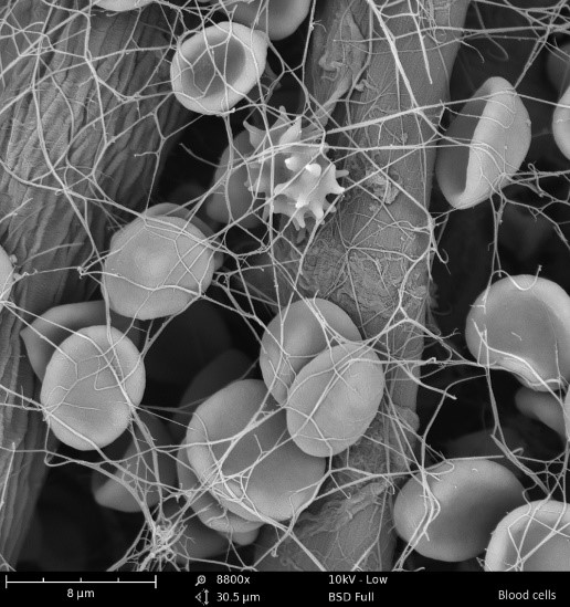
SEM produces images from a sample by scanning the surface with a focused beam of electrons in order to visualize individual cells in detail (Figure 1). These electrons can 1) excites secondary electrons deriving from the surface of the sample or 2) they can be reflected directly (backscattered) from the sample. Detectors collect the secondary and backscattered electrons to produce an image.
SEM also provides information regarding tissue microstructure, such as bone for implantation studies (measurement and pathophysiology) to analyze tissue response to various classes of implanted biomaterials.
Inorganic crystals and a wide variety of synthetic and natural materials are easily imaged using our SEM, which can be further characterized using EDX.
EDX
Our SEM system is equipped with an energy-dispersive X-ray spectroscopy which detects X-ray produced by the interaction of the electrons with the sample. Analysis of the X-ray signals are used to map the distribution and estimate the abundance of specific elements in the analyzed sample.
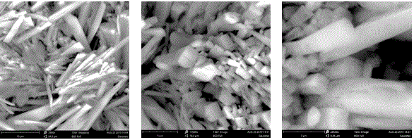
For instance, EDX identifies and characterizes metal or salt aggregates (Figure 2 and 3) within a defined area or at a specific point within the sample for toxicology studies.

Red blood cells, a macrophage and strands of fibrin inside a vessel. Magnification 8800X, scale bar image 8µm
(Image courtesy of Thermo Fisher Scientific)

Crystal aggregates of calcium phosphates (element identification using EDX). Left image: Magnification 7800X, scale bar image 10µm. Central image: Magnification 14500X, scale bar image 5µm. Right image: Magnification 28500X, scale bar image 3µm

Chemical Composition analysis of calcium phosphates crystal via EDX. A selected region was defined for the analysis. The software indicates calculate the percentage of individual identified element (top right). EDX spectrum is also shown with peaks for the identified elements.

